Pediatric Laryngospasm: Recognition, Triggers, and In-Office Rescue Protocols
 Sedation and Anesthesia Education
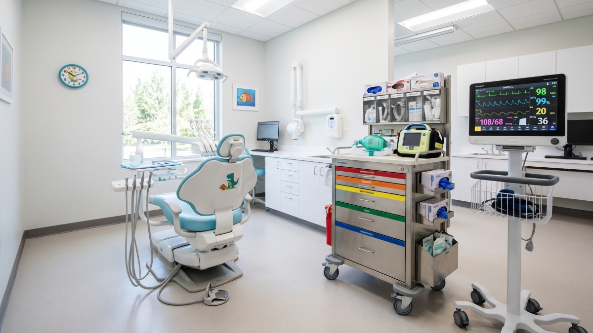
Managing a pediatric patient under procedural sedation requires constant vigilance, especially when working directly within the airway. Pediatric dental sedation introduces inherent challenges: the oral cavity doubles as the surgical field and the primary conduit for respiration. Among the most urgent emergencies a dental team can encounter is acute laryngospasm.
Because pediatric patients possess distinct anatomical vulnerabilities and lower physiological reserves compared to adults, mild respiratory compromise can escalate to severe hypoxia and bradycardia within seconds. Establishing a systematic approach to risk assessment, immediate clinical recognition, and chairside rescue protocols is essential for every practice providing pediatric sedation.
𝗪𝗵𝘆 𝗣𝗲𝗱𝗶𝗮𝘁𝗿𝗶𝗰 𝗣𝗮𝘁𝗶𝗲𝗻𝘁𝘀 𝗔𝗿𝗲 𝗮𝘁 𝗘𝗹𝗲𝘃𝗮𝘁𝗲𝗱 𝗥𝗶𝘀𝗸
Pediatric airway anatomy differs fundamentally from adult anatomy. Children under six years of age represent the highest-risk demographic for laryngospasm and adverse airway events.
Key anatomical and physiological factors include:
- 𝗟𝗮𝗿𝗴𝗲 𝗧𝗼𝗻𝗴𝘂𝗲 𝗮𝗻𝗱 𝗖𝗼𝗺𝗽𝗹𝗶𝗮𝗻𝘁 𝗔𝗶𝗿𝘄𝗮𝘆: A relatively large tongue easily impinges on the soft palate, while a softer, highly compliant trachea is prone to collapse during dynamic airway maneuvers.
- 𝗛𝘆𝗽𝗲𝗿𝘁𝗿𝗼𝗽𝗵𝗶𝗰 𝗟𝘆𝗺𝗽𝗵𝗼𝗶𝗱 𝗧𝗶𝘀𝘀𝘂𝗲: Tonsillar and adenoidal hypertrophy (Brodsky Class 3 or 4) significantly narrows the pharyngeal space and increases baseline airway resistance.
- 𝗥𝗮𝗽𝗶𝗱 𝗗𝗲𝘀𝗮𝘁𝘂𝗿𝗮𝘁𝗶𝗼𝗻: Pediatric patients exhibit a higher baseline metabolic rate and higher oxygen consumption. During complete airway obstruction or apnea, a child can desaturate to critical hypoxia levels in roughly half the time it takes a healthy adult.
𝗜𝗱𝗲𝗻𝘁𝗶𝗳𝘆𝗶𝗻𝗴 𝗖𝗹𝗶𝗻𝗶𝗰𝗮𝗹 𝗧𝗿𝗶𝗴𝗴𝗲𝗿𝘀
Laryngospasm is a protective reflex closure of the vocal cords that becomes exaggerated and pathologic. In the dental operatory, several factors precipitate this event:
- 𝗙𝗼𝗿𝗲𝗶𝗴𝗻 𝗠𝗮𝘁𝘁𝗲𝗿 𝗶𝗻 𝘁𝗵𝗲 𝗣𝗵𝗮𝗿𝘆𝗻𝘅: Water spray, blood, prophy paste, or pooled secretions touching the vocal cords can immediately trigger involuntary cord closure.
- 𝗟𝗶𝗴𝗵𝘁 𝗣𝗹𝗮𝗻𝗲𝘀 𝗼𝗳 𝗦𝗲𝗱𝗮𝘁𝗶𝗼𝗻: Involuntary protective airway reflexes are hyperactive during lighter stages of sedation. Procedures initiated during transitional depths are particularly vulnerable.
- 𝗔𝗶𝗿𝘄𝗮𝘆 𝗛𝘆𝗽𝗲𝗿𝗿𝗲𝗮𝗰𝘁𝗶𝘃𝗶𝘁𝘆: Patients presenting with a recent upper respiratory infection (URI), active allergies, or secondhand smoke exposure have sensitized, hyperreactive vocal cords. If a child presents with wet lung sounds, rhonchi, or active congestion, the safest decision is to reappoint the procedure.
𝗥𝗲𝗰𝗼𝗴𝗻𝗶𝘁𝗶𝗼𝗻: 𝗣𝗮𝗿𝘁𝗶𝗮𝗹 𝘃𝘀. 𝗖𝗼𝗺𝗽𝗹𝗲𝘁𝗲 𝗦𝗽𝗮𝘀𝗺
Rapid recognition is crucial before pulseless arrest or severe neurological compromise occurs. Practitioners should recognize the two distinct presentations:
- 𝗣𝗮𝗿𝘁𝗶𝗮𝗹 𝗟𝗮𝗿𝘆𝗻𝗴𝗼𝘀𝗽𝗮𝘀𝗺: High-pitched, inspiratory stridor or "crowing" sounds accompany labored chest wall excursion. Some gas exchange remains, but airflow is severely restricted.
- 𝗖𝗼𝗺𝗽𝗹𝗲𝘁𝗲 𝗟𝗮𝗿𝘆𝗻𝗴𝗼𝘀𝗽𝗮𝘀𝗺: Characterized by absolute silence. The patient will demonstrate paradoxical chest wall and abdominal movement (see-saw breathing) and tracheal tugging without actual ventilation.
𝗖𝗹𝗶𝗻𝗶𝗰𝗮𝗹 𝗣𝗲𝗮𝗿𝗹: Never mistake silence for normal breathing. A silent chest with vigorous respiratory effort is a hallmark of complete upper airway obstruction or total laryngospasm.
𝗜𝗻-𝗢𝗳𝗳𝗶𝗰𝗲 𝗥𝗲𝘀𝗰𝘂𝗲 𝗣𝗿𝗼𝘁𝗼𝗰𝗼𝗹
When laryngospasm occurs, the dental team must act decisively through a graduated escalation pathway:
- 𝗖𝗲𝗮𝘀𝗲 𝗧𝗿𝗲𝗮𝘁𝗺𝗲𝗻𝘁 𝗮𝗻𝗱 𝗖𝗹𝗲𝗮𝗿 𝘁𝗵𝗲 𝗙𝗶𝗲𝗹𝗱: Immediately stop the procedure, pack the surgical site if bleeding, and thoroughly suction the oral cavity to eliminate continuing mechanical stimuli.
- 𝗔𝗽𝗽𝗹𝘆 𝟭𝟬𝟬% 𝗣𝗼𝘀𝗶𝘁𝗶𝘃𝗲 𝗣𝗿𝗲𝘀𝘀𝘂𝗿𝗲 𝗢𝘅𝘆𝗴𝗲𝗻: Fit a tight-fitting mask to the child's face and apply gentle positive pressure ventilation using 100% O2 via a bag-valve-mask (BVM) system.
- 𝗘𝘅𝗲𝗰𝘂𝘁𝗲 𝘁𝗵𝗲 𝗟𝗮𝗿𝘀𝗼𝗻 𝗠𝗮𝗻𝗲𝘂𝘃𝗲𝗿: Apply firm, inward and anterior pressure bilaterally at the "laryngospasm notch" (located behind the ramus of the mandible, between the mastoid process and the condyle) combined with a vigorous jaw thrust. This painful stimulus frequently breaks the reflex arc and opens the glottis.
- 𝗣𝗵𝗮𝗿𝗺𝗮𝗰𝗼𝗹𝗼𝗴𝗶𝗰 𝗥𝗲𝘀𝗰𝘂𝗲: If the spasm does not break and desaturation continues, emergency neuromuscular blockade (such as succinylcholine, paired with atropine to counter vagal bradycardia) must be administered via IV, IM, or intraosseous routes by trained personnel.
𝗦𝘁𝗮𝗻𝗱𝗮𝗿𝗱𝗶𝘇𝗶𝗻𝗴 𝗣𝗿𝗮𝗰𝘁𝗶𝗰𝗲 𝗣𝗿𝗲𝗽𝗮𝗿𝗲𝗱𝗻𝗲𝘀𝘀
Sedation safety is built on structured pre-operative evaluation, strict NPO adherence, continuous clinical monitoring, and comprehensive emergency readiness.
To refresh your practice protocols, update your sedation emergency systems, and meet state compliance standards, visit https://www.isedatesafe.com. Ensuring your entire team conducts regular in-office airway drills transforms unexpected clinical complications into manageable, predictable events.
Categories: : Patient Safety, Sedation Anesthesia